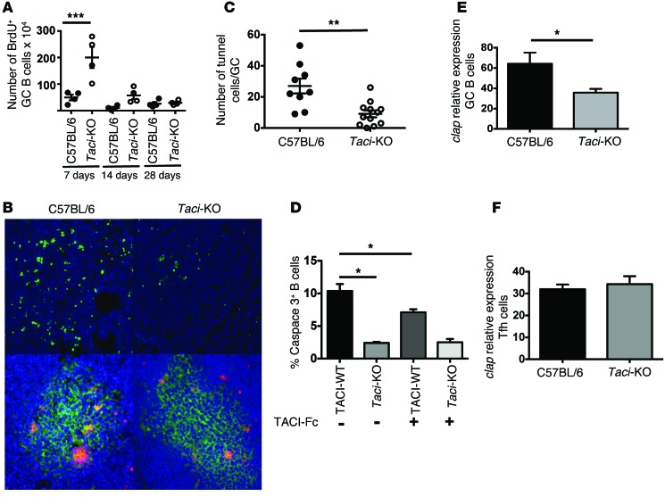
Figure 3. Impact of TACI deficiency on B cell proliferation and apoptosis.
(A) Number of proliferating GC B cells in WT and TACI-deficient mice at 7, 14, or 28 days after immunization with SRBCs. Percentages of BrdU+ GC cells (CD19+GL7+FAS+). TACI deficiency increased proliferation of GC cells 7 days after immunization (P = 0.0002, Holm-Sidak test). (B) Immunofluorescence analysis of GC cell apoptosis. Apoptotic cells were identified by TUNEL, labeled ends were detected with anti-BrdU antibodies (green, upper diagrams and red, lower diagrams), and cells were counterstained with DAPI. Lower diagrams show costaining with anti-GL7 mAbs (green). Results are representative of 6 distinct sections obtained from 2 WT mice and 3 TACI-deficient mice. Original magnification, ×200. (C) Enumeration of apoptotic TUNEL+ GC cells. Taci-KO mice had fewer TUNEL+ cells per GC (P = 0.0012, Mann-Whitney U test). (D) Impact of TACI ligand blockade on B cell apoptosis. WT or Taci-KO splenic B cells were cultured with anti-CD40 and IL-4 in the presence or absence of TACI-Fc to inhibit TACI signaling. After 48 hours, caspase 3+ cells were enumerated by FACS. TACI deficiency or impaired TACI function decreased apoptosis after B cell activation, (P < 0.05, Mann-Whitney U test). (E and F) Relative expression by qPCR of cellular inhibitor of apoptosis (cIap) by GC B cells (E) or Tfh cells (F) 14 days after immunization with SRBCs. Expression of cIap was quantified in cDNA by qPCR reactions using specific primers and SYBR Green incorporation (P < 0.05, Mann-Whitney U test).

