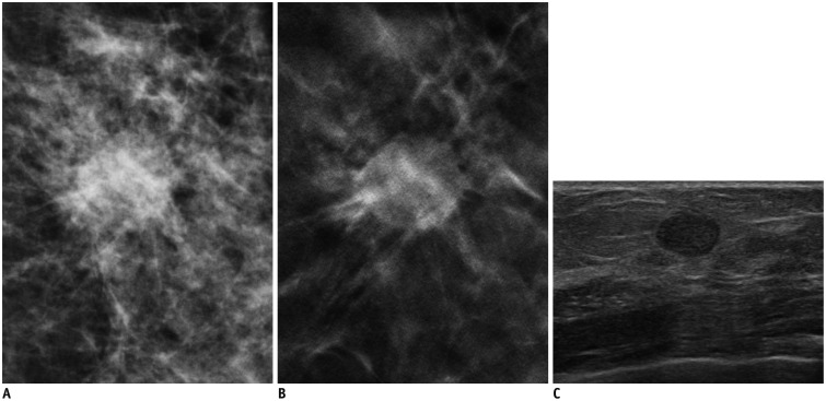
Fig. 2. 57-year-old woman with 1.2 cm-sized invasive papillary carcinoma.
A. Craniocaudal full-field digital mammography view. B. Craniocaudal digital breast tomosynthesis (DBT) (1-mm section) view. C. Transverse ultrasonography (US) view. Mass with spiculated margin is clearly visible on DBT image. This lesion appears as oval circumscribed lesion on US. Readers 1 and 2 categorized this lesion as Breast Imaging Reporting and Data System (BI-RADS) category 4B, and reader 3 categorized this lesion as BI-RADS category 4A on DBT. Readers 2 and 3 categorized this lesion as BI-RADS category 3, and reader 1 categorized this lesion as BI-RADS category 4A on US.

