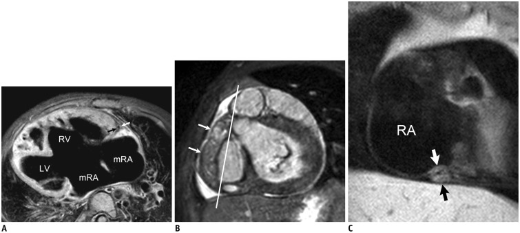
Fig. 6. Coronary vessel wall MRI.
A. Axial electrocardiography (ECG)-triggered, navigator-gated, double inversion recovery, fat-saturated black-blood segmented turbo spin-echo MRI in 3-year-old girl with functional single ventricle and right isomerism who underwent bidirectional cavopulmonary shunt shows wall (arrows) of normal right coronary artery. In addition, dextrocardia, large ventricular septal defect, and remnant of interatrial septum are noted. C. Oblique coronal ECG-triggered, navigator-gated, double inversion recovery, fat-saturated black-blood segmented turbo spin-echo MRI obtained along line in B in 3-year-old boy with Kawasaki disease and thrombosed fusiform aneurysm (long arrows in B) of right coronary artery reveals severe concentric wall thickening (short arrows in C) of distal right coronary artery. LV = left ventricle, mRA = morphologic right atrium, RA = right atrium, RV = right ventricle

