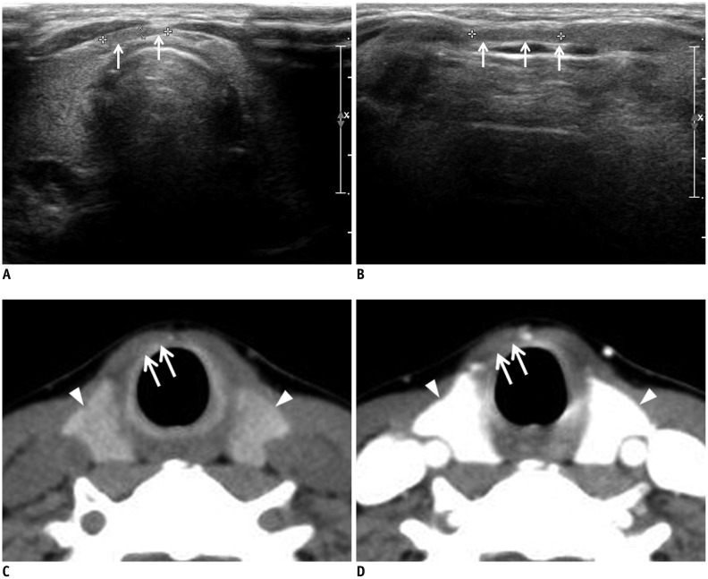
Fig. 2. Fatty tissue mimicking thyroid pyramidal lobe on ultrasonography in 49-year-old woman.
Transverse (A) and longitudinal (B) gray-scale sonograms show longitudinally arranged structure mimicking thyroid pyramidal lobe (arrows). Nonenhanced (C) and contrast-enhanced (D) axial CT images shows only fatty tissue in same position (arrows), and they show different attenuation and enhancement from main thyroid gland (arrowheads).

