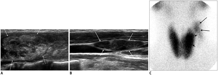
Fig. 12. Papillary thyroid cancer presenting with hyperthyroidism in 12-year-old female.
Longitudinal ultrasonography scan (A) shows oval hyperechoic nodule with numerous microcalcifications (arrows) and multiple metastatic lymphadenopathy in left thyroid at levels II-IV (B). Microcalcifications and cystic changes are seen. C. Tc-99m scintigraphy shows increased uptake (thyroid uptake 7.2%; normal range, 2-4%), diffuse enlargement of both thyroid gland lobes, and increased uptake in thyroid nodule (short arrows) and in left lateral neck due to metastasis to lymph nodes (long arrows). Surgery confirmed papillary thyroid cancer with metastatic lymphadenopathy and underlying lymphocytic thyroiditis.

