Figure 7.
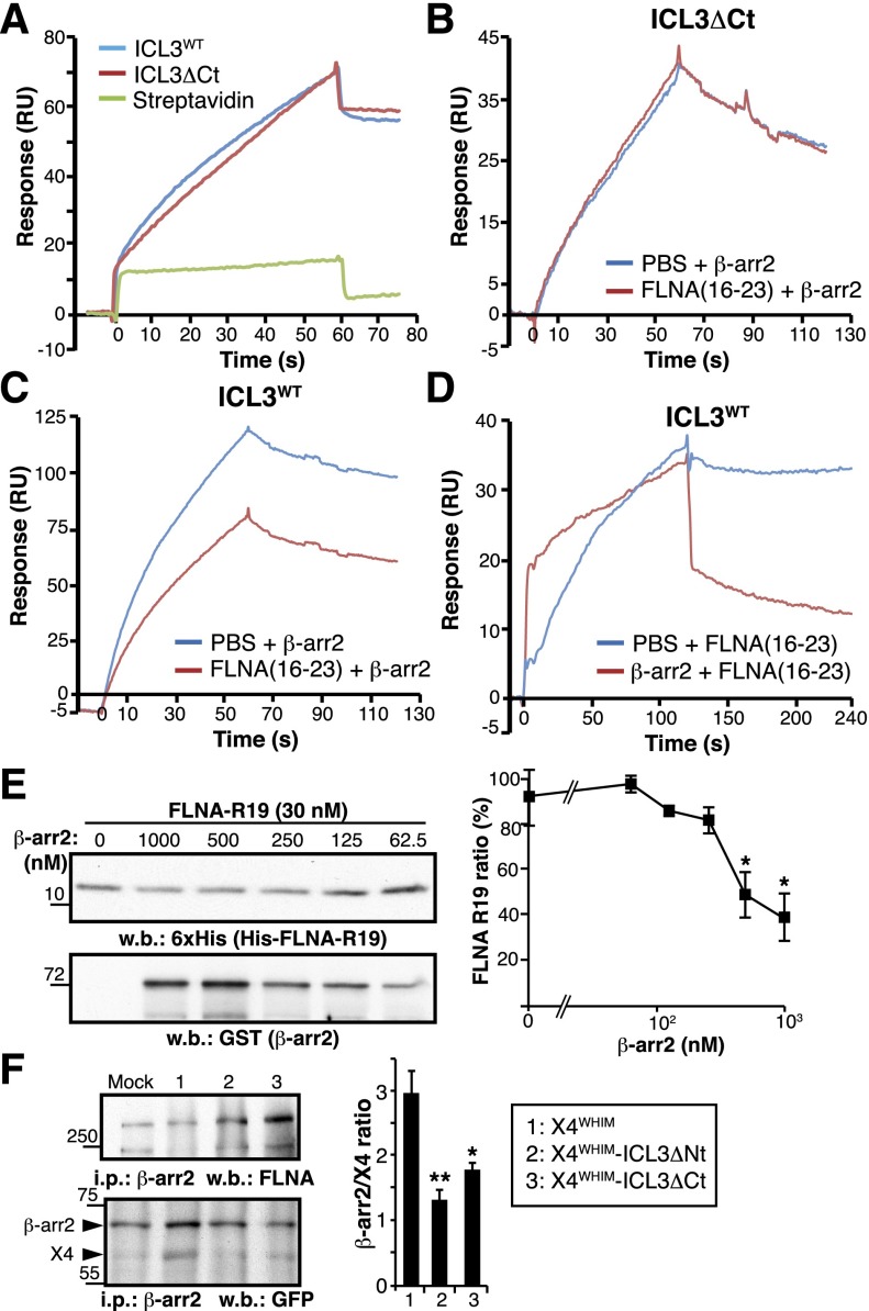
FLNA-ICL3 interaction enhances β-arr2 association with WHIM receptor in cells. (A) BiaCore sensorgrams for β-arr2 binding to immobilized ICL3, ICL3ΔCt peptides, and streptavidin (reference), monitored as RU. The β-arr2 concentration was 20 nM in all cases. (B-C) SPR analysis of FLNA interference with β-arr2 binding to ICL3. The FLNA(16-23) fragment (400 nM) and β-arr2 (20 nM) were injected sequentially onto flow cells with immobilized (B) ICL3ΔCt or (C) ICL3WT peptides. Curves depict β-arr2 binding (in RU) to each peptide after subtraction of the RU signal for FLNA(16-23) fragment binding. (D) Analysis of simultaneous β-arr2 and FLNA binding to ICL3. β-arr2 (200 nM; red curve) or buffer (blue curve) were injected onto ICL3WT, followed by injection of the FLNA(16-23) fragment (250 nM). The sensorgrams show the association and dissociation phases of the FLNA(16-23) fragment in each condition, after subtraction of β-arr2 and buffer RU values. (E) Analysis of FLNA interference with β-arr2 binding to ICL3 by pull-down assay with purified His-tagged FLNA repeat 19 fragment and glutathione S-transferase (GST) –β-arr2 at indicated concentrations. Representative immunoblots are shown with anti-6xHis and anti-GST antibodies and densitometric quantification. (F) Analysis of FLNA and β-arr2 binding to X4WHIM receptor variants in living cells. GFP-tagged X4WHIM, X4WHIM-ΔNt, and X4WHIM-ΔCt receptors were transfected in HEK-293 cells stably expressing GFP-β-arr2. Equal amounts of cell lysates were immunoprecipitated with anti-β-arr2 antibody, and the precipitated proteins were analyzed with anti-FLNA and anti-GFP antibodies. Bands corresponding to β-arr2 and CXCR4 were quantified by densitometry (right), and the ratio was calculated by using the values for mock cells as reference. (E-F) Data are mean ± SEM (n = 3). **P < .02; *P < .05, 1-tailed Student t test compared with X4WHIM cells.

