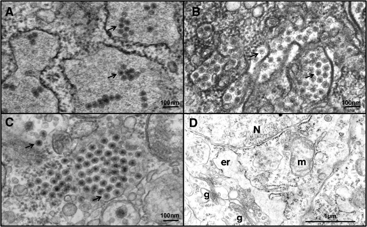
Figure 2.
Transmission electron microscopy photographs showing evidence of viral infection in C6/36 cell cultures inoculated with homogenates of laboratory colony mosquitoes. (A) Aedes aegypti-Galveston and (B) Aedes albopictus-Thailand with arrows indicating flavivirus virions, and (C) Aedes albopictus-Galveston with arrows indicating reovirus-like structures, and (D) ultrastructure of uninfected C6/36 cells in culture. N = nucleus, er = swollen cistern of granular endoplasmic reticulum, g = Golgi apparatus, m mitochondrion.

