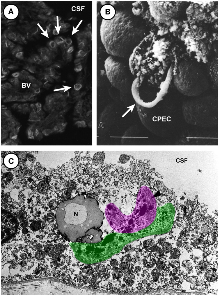
Figure 4.
Biondi ring tangles in aged human choroid plexus of the lateral and fourth choroid plexus. (A) Fluorescent light micrograph of thioflavin S-stained Biondi ring tangles (arrows) in the choroid plexus of the brain of a 78-year-old human female with Alzheimer's disease showing Biondi ring tangles appear as ring, tangle, serpentine, and curled profiles. (Magnification: 530×). (B) Scanning electron micrograph (Magnification 2500 ×) showing destruction of plexus epithelial cell containing ring-like Biondi inclusions. Arrow marks a ring bursting from an individual plexus epithelial cell. Material from 78 year old woman. (C) Electron micrograph of a choroid plexus from a 70-year-old male with Alzheimer's disease showing the fibrous Biondi ring tangles (one highlighted in pink, the other in green) associated with lipofuscin granules, mitochondria, and other cellular components. (Magnification: 10,300×). (A,C) Reproduced from Wen et al., 1999 Copyright © 1999 Elsevier Science B.V. All rights reserved. (B) Reproduced from (Kiktenko, 1986) Copyright © 1986 Springer All rights reserved. Abbreviations: BV, blood vessel; CPEC, choroid plexus epithelial cell; CSF, cerebrospinal fluid; N, nucleus.

