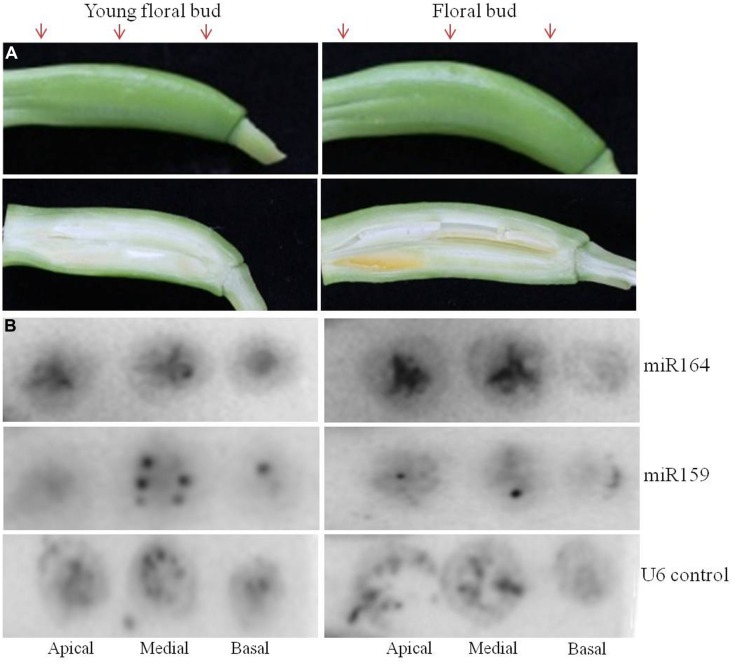
FIGURE 4.
Spatio-temporal expression of miR164 and miR159 by tissue printing hybridization in floral buds of agave. Expression of miR164 and miR159 was analyzed in the apical, medial, and basal region of agave floral buds by tissue printing hybridization. Two different stages of agave floral buds were analyzed. (A) Transverse sections of the agave floral buds were used to make the tissue prints; arrows indicate the apical, medial, and basal region were the cuts were made. Below each floral bud a longitudinal section is shown to indicate the developmental stage. (B) Detection of miR164, miR159, and nucleolar U6 (positive control) in agave floral buds by hybridization.

