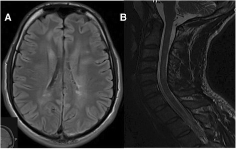
Figure 1.

MRI brain and cervical spine images. A) Axial Fluid-Attenuated Inversion Recovery (FLAIR) image of the brain showing demyelinating plaques in periventricular and juxtacortical regions; B) Sagittal T2 image of the cervical spine showing intramedullary demyelinating plaques at C3 and C4-C5 levels.
