Figure 3.
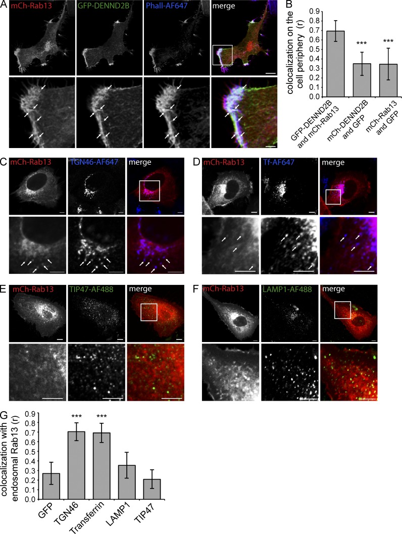
Rab13 colocalizes with DENND2B at the cell’s leading edge. (A) MCF10A cells were cotransfected with GFP-DENND2B and mCh-Rab13, fixed, and stained with Phalloidin–Alexa Fluor 647 (AF647). Bars: (low magnification) 10 µm; (high magnification) 2.5 µm. (B) Quantification of colocalization at the cell periphery using Pearson correlation coefficient (mean ± SD measuring 10 cells per condition pooled from two independent experiments; one-way ANOVA with Dunnett’s post-test; ***, P < 0.001). (C–F) MCF10A expressing mCh-Rab13 were fixed and labeled with TGN46-AF647 (C) internalized transferrin-AF647 (D), TIP47-AF488 (E), or LAMP-1–AF488 (F). Arrows point to colocalizing structures. Bars, 5 µm. (G) Quantification of colocalization from C–F as described in B. Boxed regions are magnified on the bottom.

