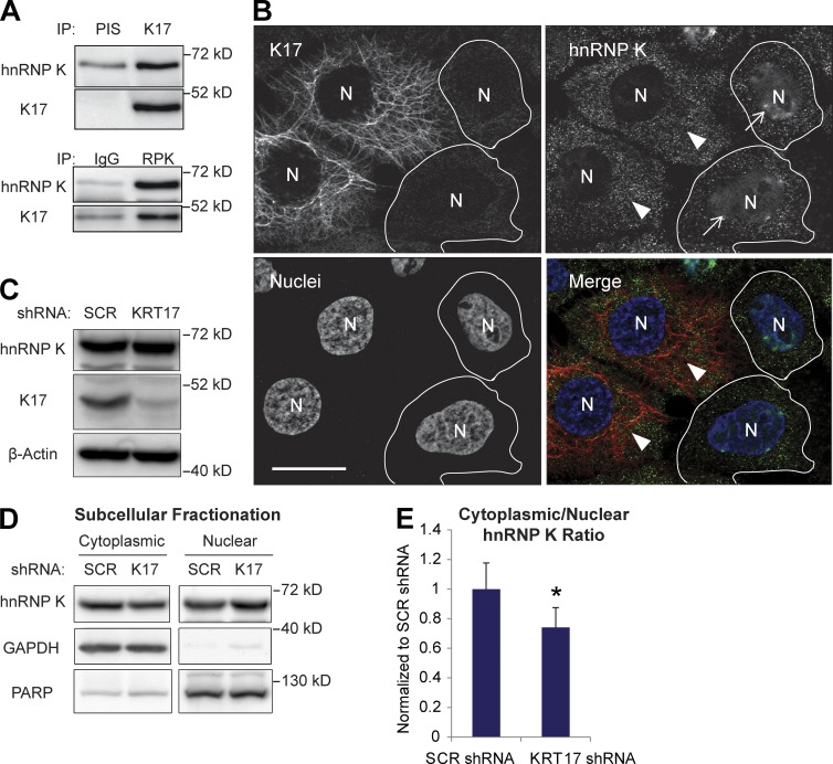
Figure 1.
K17 binds to hnRNP K and regulates cytoplasmic localization of hnRNP K. (A) Co-IP of K17 and hnRNP K (RPK). IP was performed in A431 cells with anti-K17 or hnRNP K (RPK) antibody with preimmune serum (PIS) or IgG as a control. Immunoblotting was performed with antibodies against the indicated proteins. (B) Immunostaining of hnRNP K (green) and K17 (red) in A431 cells transduced with KRT17 shRNA. Nuclei (N) are shown in blue. Arrows indicate cells expressing KRT17 shRNA and arrowheads indicate cells (outlined) that did not become transduced with KRT17 shRNA. Bar, 10 µm. (C) Cell lysates from A431 cells stably expressing SCR or KRT17 shRNA were subjected to SDS-PAGE and immunoblotting was performed with the antibodies against the indicated proteins. (D) Subcellular fractionation of hnRNP K in A431 cells stably expressing SCR or KRT17 shRNA. Immunoblotting was performed with antibodies against the indicated proteins. PARP was used as a control for the nuclear fraction, whereas GAPDH was used for the cytoplasmic fraction. (E) Signal intensities of hnRNP K bands from D were quantitated and shown as the cytoplasmic-to-nuclear hnRNP K ratio. Data from three experimental repeats were normalized to A431 SCR shRNA cells and are represented as mean ± SEM (error bars). *, P < 0.03.

