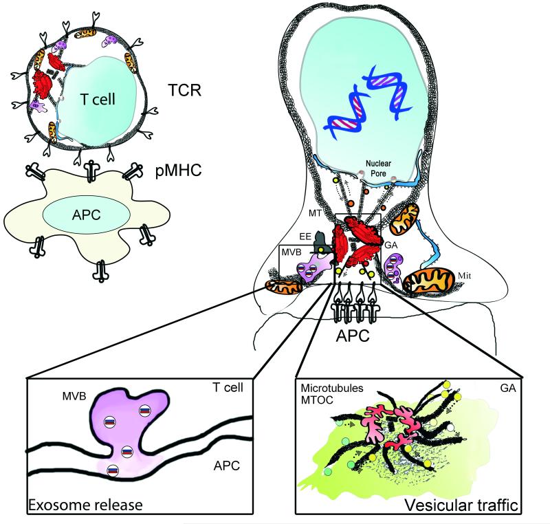
Figure 1. Polarization of organelles at the immune synapse.
The IS forms rapidly upon focal TCR activation. The TCR accumulates at the interface of the T cell and the stimulating surface; an antigen-presenting cell (APC) or a surface coated with stimulating antibodies or recombinant proteins, such as the MHC complex. The centrosome localizes to the IS and brings the associated Golgi Apparatus (GA). The early endosome compartment (EE) regulates recycling of surface receptors. The mitochondrial network (Mit), in association with the endoplasmic reticulum, dynamically localizes to the IS and provides a controlled calcium flux and a focused supply of ATP for energy provision. Nuclear activating molecules enter the nucleus via nuclear pores. They can be transported in vesicles or through direct interaction with molecular motors such as Dynein. Left inset: Multivesicular bodies (MVB) localize to the IS upon centrosome relocalization and have several functions, including the recycling of proteins and the formation of exosomes. MVBs serve as exocytosis compartments at the IS. Right inset: vesicular traffic at the IS. Vesicles coming from the IS are directed to recycling organelles, such as endosomes. While a number of vesicles emerging from the trans-Golgi network facilitate polarized secretion.

