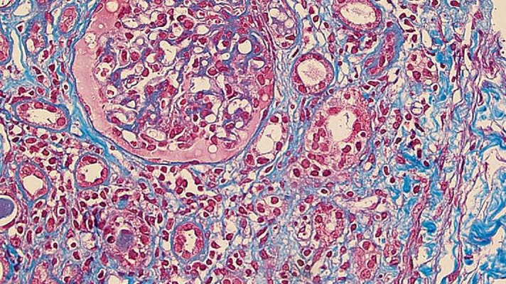
Figure 1.

The renal biopsy specimen. This panel shows a high-power image (×400) of a slide prepared from the renal biopsy specimen (Azan and Masson trichrome stain). The optical microscopic study showed almost normal glomeruli and diffuse infiltration of mononuclear cells in the interstitium. The mononuclear cells were mostly lymphocytes, with a few plasma cells. Furthermore, this panel shows that mononuclear cells were infiltrating the tubules. A low-power image (×20) revealed that fibrosis was present in 30% of the whole tissue.
