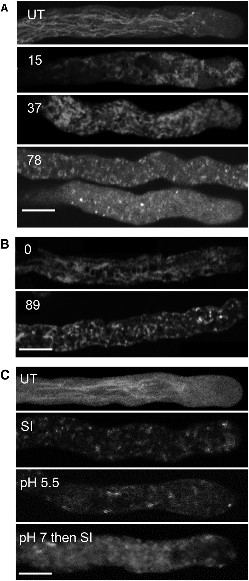
Figure 6.
SI triggers pollen tube vacuolar reorganization and disintegration, and artificial acidification of the cytosol triggers alterations in vacuolar organization. A, Pollen tubes labeled with 1 µm carboxy-DCFDA were SI induced and imaged using confocal microscopy. Numbers indicate the length of treatment time in minutes: t = 15 min indicates that the vacuolar network has reorganized, t = 37 min indicates additional vacuolar reorganization, and t = 78 min indicates that the vacuolar structure appears disintegrated. UT, Typical untreated pollen tube with a tubular vacuole organization. B, Stills from a time series of a pollen tube expressing δ-TIP-GFP: t = 0 indicates typical membrane-localized reticulate structure, and t = 89 min indicates that, after SI induction, there is apparent reorganization and only a dispersed, punctate signal remains. C, Field poppy pollen tubes were labeled with carboxy-DCFDA and imaged using confocal microscopy after the following treatments: UT, Untreated; SI, SI-induced pollen tube for t = 76 min; pH 5.5, 50 mm propionic acid (pH 5.5)-treated pollen tube for t = 85 min; pH 7 then SI, pollen tube pretreated with 50 mm propionic acid (pH 7.0) for t = 10 min followed by SI induction for t = 68 min. Bars = 10 µm.

