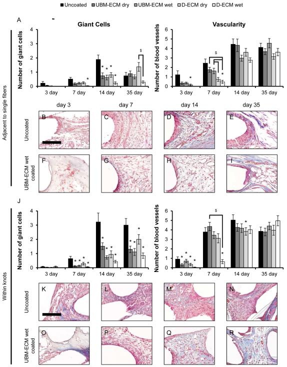
Figure 8.
Histologic remodeling response to uncoated and ECM hydrogel coated mesh. The total number of foreign body giant cells and blood vessels were quantified from high magnification (400X) Masson’s Trichrome stained images adjacent to single mesh fibers (A) and within mesh fiber knots (J) after 3, 7, 14, and 35 days post implantation. Representative images of uncoated polypropylene mesh (B-E, K-N) and UBM-ECM wet coated mesh (F-I, O-R) are shown at each time point. Statistical significant differences were determined with ANOVA (p < 0.05) and denoted with (*) as different from uncoated mesh, or ($) as different between ECM coating groups. Scale bar represents100 μm.

