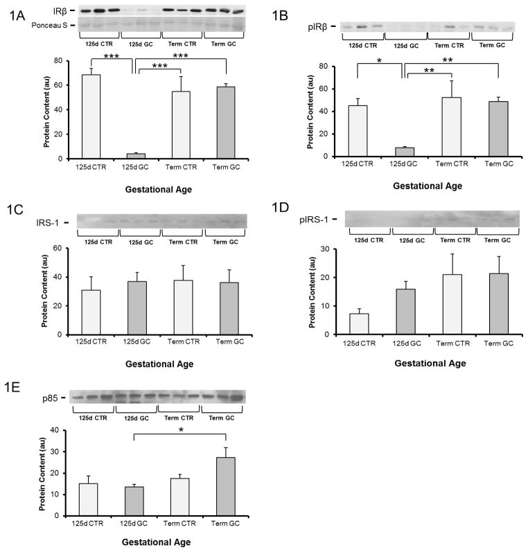
Figure 1. Effect of glucocorticoid treatment (GC) on insulin signaling protein content and phosphorylation in muscle from fetal baboons at 0.67 gestation and term baboons.
Insulin receptor (IR)-β (A), IR-β Tyr 1361 phosphorylation (pIR-β) (B), IR substrate-1 (IRS-1) (C), IRS-1 tyrosine phosphorylation (pIRS-1) (D), and p85 subunit of PI 3-kinase (p85) (E) were measured by Western blotting and immunoprecipitation. n=5–6 per group. Representative blots from 3 animals per group are also shown. d-day; CTR-control; GC-glucocorticoid. Graphical data are means ± SE. *p<0.05, **p<0.01, ***p<0.001.

