Fig. 3.
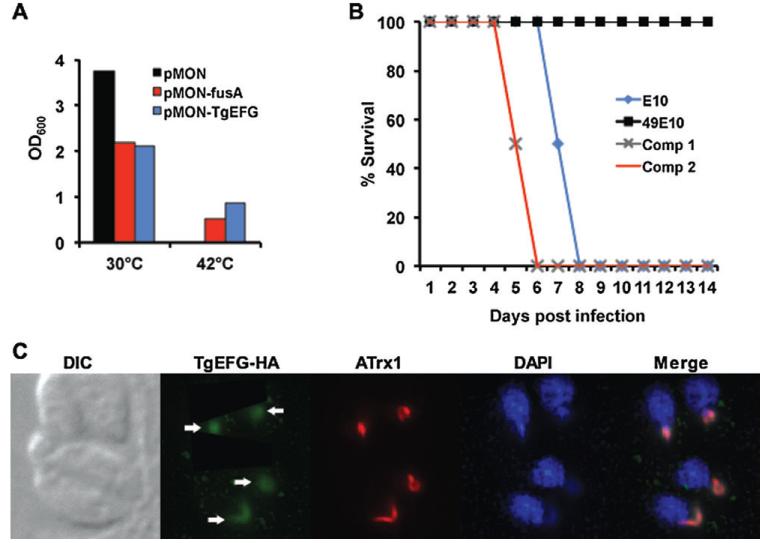
TgEFG functions as EFG, restores virulence to 49E10 and is localized to the apicoplast
A. A temperature-sensitive EFG mutant strain of E. coli was transfected with either vector alone (pMON, black bar), the vector expressing EFG from E. coli (pMON-fusA, red bar), or the vector expressing TGME49_02370 from T. gondii (pMON-TgEFG, blue bar). All cells were grown at the permissive temperature (30°C) to similar turbidity corresponding to mid-log phase and equivalently diluted into fresh broth containing 1mM IPTG. Identical cultures were grown for 16 hours at both the permissive (30°C) and restrictive (42°C) temperatures. OD600 measurements as an indictor of growth are shown.
B. The virulence of E10 (blue diamond) and 49E10 (black square) parasites was compared to two independent clones of 49E10 complemented with TgEFG expressed from its endogeneous promoter (Comp 1 grey X and Comp 2 red line). For this acute mouse model, 1.5×10⁶ tachyzoites were i.p. injected into four mice per strain. Two independent experiments were combined for a total of eight mice per strain.
C. The cellular localization of TgEFG was examined by IFA using rabbit anti-HA antibody to visualize HA-tagged TgEFG expressed from its endogenous promoter (green), the 11G8 monoclonal to stain ATrx1 (red) and DAPI to visualize nucleic acid (blue). Fluorescent images were acquired as previously described (Mordue et al., 2007). White arrows indicate the TgEFG-HA in the apicoplast.

