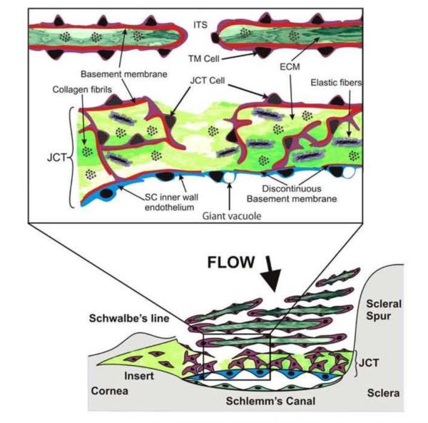
Figure 1.
Diagram of the outflow pathway and juxtacanalicular (JCT) or cribriform region. The lower portion of the figure shows a stylized view of the TM and the upper inset shows an expanded view of the JCT region. TM = trabecular meshwork, ECM = extracellular matrix, SC = Schlemm’s canal, ITS = intertrabecular space
Reprinted with permission from Acott TS, Kelley MJ Extracellular matrix in the trabecular meshwork. Exp Eye Res (2008) 86: 543-561.

