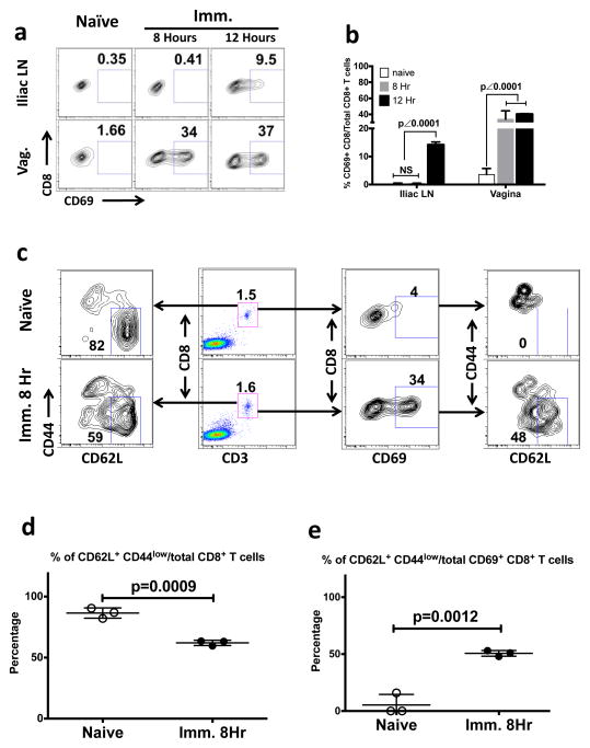
Figure 5. The local immune priming of endogenous CD8+ T cells that naturally migrate into the physiologically intact vaginal mucosa.
The phenotype of vaginal CD8+ T cells in the Ivag SIINFEKL-immunized RAG-I KO, OT-I transgenic mice. This is representative of 2 independently repeated experiments (n=3) with the same results. (a and b) The CD69 expression of CD8+ T cells in vaginal mucosa and iliac LNs of naïve and immunized animals that were sacrificed at 8 hours and 12 hours PI. (a) Gating strategy. (b) The statistical difference (mean ± SEM) among naïve and 2 immunized groups. The p value was generated using a one-way ANOVA plus Tukey’s multiple comparison. (c) The gating strategies for the phenotype assay of vaginal CD8+ T cells in naïve and immunized animals at 8 hours after immunization. Left plots, The percentage of CD62L+ and CD44low cells out of total vaginal CD8+ T cells. Middle left plots, The percentage of vaginal CD8+ T cells. Middle right plots, The percentage of CD69 expressing cells out of total vaginal CD8+ T cells. Right plots, The percentage of CD62L+ CD44low cells out of total CD69 expressing vaginal CD8+ T cells. (d) The statistical difference (mean ± SEM) of panel C, left plots. (e) The statistical difference (mean ± SEM) of panel C, right plots. The p values were generated using Student’s t test.

