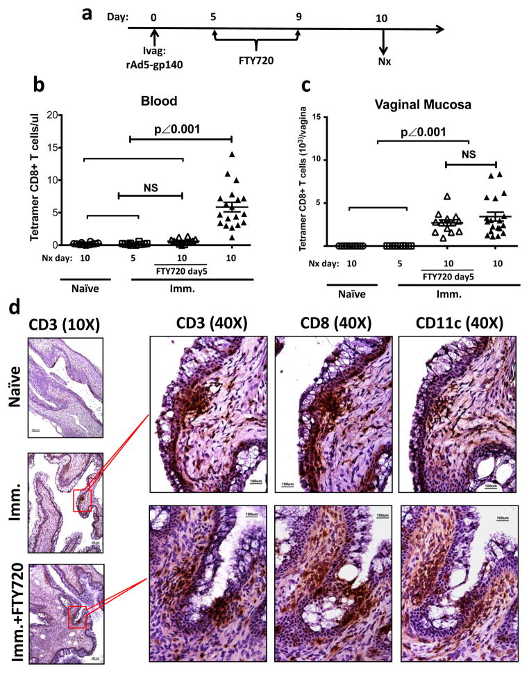
Figure 7. Local formation of Ag-specific IVALT in the vaginal mucosa of Ivag immunized WT mice.
(a to c) The absolute number of Ag-specific CD8+ T cells in blood and vaginal mucosa of naïve and immunized mice either with or without FTY720 treatment. ○, naïve mice euthanized at day 10 PI (n=12); □, immunized mice euthanized at day 5 PI (n=12); △, FTY720 treated immunized mice euthanized at day 10 PI (n=12); ▲, immunized mice euthanized at day 10 PI (n=18). This is the pool of 3 independently repeated experiments with the same results. (a) The experimental design. (b) The absolute number (mean ± SEM) of Ag-specific CD8+ T cell in blood. (c) The absolute number (mean ± SEM) of Ag-specific CD8+ T cell in vaginal mucosa. The p values were generated using one-way ANOVA plus Tukey’s multiple comparison. (d) Representative IHC staining showing the formation of immune cell clusters containing CD3+, CD8+ and CD11c+ cells in immunized mice either with or without FTY720 treatment. Scale bar: 100 μm in 40X photomicrographs; 400 μm in 10X photomicrographs.

