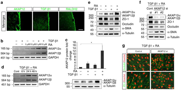
Figure 2. A combination of TGF-β1 and RA induces high levels of AKAP12 expression, which maintains the epithelial properties of the meninges.

(a) AKAP12 expression was co-localized with TGF-β1 and RALDH2 in the normal meninges in vivo. Scale bar, 40 μm (upper panels), 200 μm (lower panels). (b) ARPE-19 cells were serum starved for 24 h, and then treated with TGF-β1 (10 ng ml−1) and RA (5 and 10 μM) for 24 h. AKAP12 mRNA levels were markedly increased by co-treatment of these two factors compared with those of single treatments. (c) Co-treatment with TGF-β1 and RA synergistically increased AKAP12 protein levels. ARPE-19 cells were serum starved for 24 h, and then treated with TGF-β1 (10 ng ml−1) and RA (10 μM) for 24 h (mean ± s.d., n = 4, analysis of variance followed by Tukey-Kramer test: *P = 0.006). (d) Reverse transcription PCR was performed after treatment with TGF- β1 (10 ng ml−1) and RA (10 μM). AKAP12 mRNA levels were significantly increased after 6 h and were sustained up to 48 h. Glyceraldehyde-3-phosphate dehydrogenase was used as an internal control (Cont). (e) RA blocked TGF-β1-induced EMT. ARPE-19 cells were serum starved for 24 h, and then treated with TGF-β1 (10 ng ml−1) and RA (10 μM) for 48 h. (f) The inhibitory effect of RA on TGF-β1 was abolished in the absence of AKAP12. AKAP12 siRNA-transfected ARPE-19 cells were serum starved for 24 h and incubated in the presence of TGF-β1 (10 ng ml−1) and RA (10 μM) for 48 h. (g) Culture conditions were the same as in f Cells on coverslips were fixed and immunostained with antibodies against occludin and ZO-1. Scale bar, 40 μm. Each panel represents the results from independent experiments repeated at least four times using different animals or conditions.
