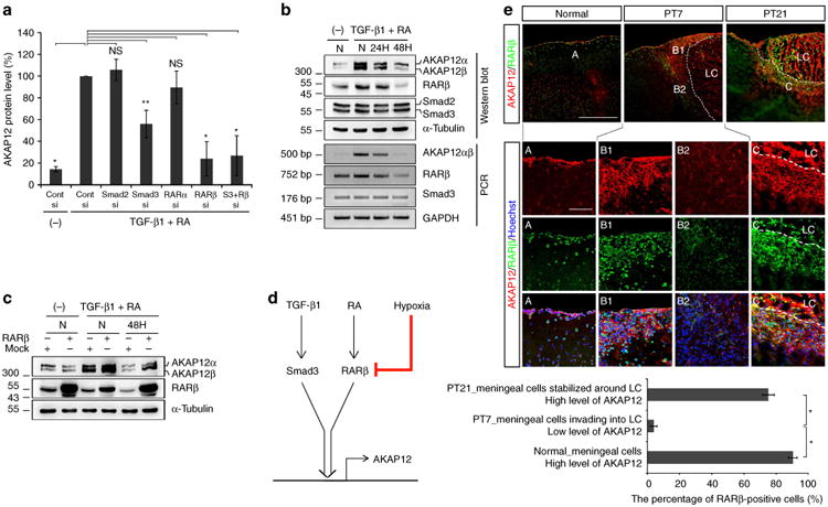
Figure 4. Oxygen tension regulates induction of AKAP12 through TGF-β1 and RA at a transcriptional level via RARβ.

(a) ARPE-19 cells were co-transfected with control (Cont), Smad2, Smad3, RARα and RARβ siRNA. Knockdown cells were treated with TGF-β1 (10 ng ml−1) and RA (10 μM) for 24 h after serum starvation for 24 h. Data are expressed relative to the control siRNA transfectant treated with TGF-β1 (10 ng ml−1) and RA (10 μM) for 24 h and are normalized to 100% (mean ± s.d. n = 3 analysis of variance (ANOVA) followed by Tukey-Kramer test: *P<0.0005 **P = 0.003; NS not significant). (b) AKAP12 expression is reduced at a transcriptional level under hypoxia and correlates with RARβ expression. ARPE-19 cells were serum starved for 24 h and incubated under normoxia or hypoxia in the presence of TGF-β1 (10 ng ml−1) and RA (10 μM). (c) RARβ overexpression recovered the reduced level of AKAP12 under hypoxia. pCMV flag human RARβ plasmid-transfected ARPE-19 cells were serum starved for 6 h and incubated under normoxia or hypoxia in the presence of TGF-β1 (10 ng ml−1) and RA (10 μM) for 48 h. (d) Diagram depicting the regulation of AKAP12 expression by hypoxia in the presence of TGF-β1 and RA. (e) Brain sections were co-stained with antibodies against AKAP12 and RARβ. AKAP12 expression has a similar pattern to that of RARβ during meningeal reconstruction. Scale bar, 500 μm (large panels), 100 μm (magnified panels). Three representative sections per mouse were analysed using Image J. Mean ± s.d., n = 4 mice per time point, ANOVA followed by Tukey-Kramer test: *P< 0.0005. N, normoxia (21% oxygen); H hypoxia (1% oxygen); LC lesion core. Panels b,c represent the results from independent experiments repeated at least four times.
