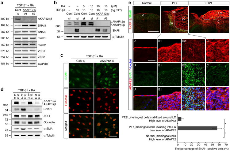
Figure 5. AKAP12 modulates EMT by regulating SNAI1 expression.

(a) AKAP12 knockdown specifically upregulated SNAI1 expression among all the EMT-inducing transcription factors analysed. ARPE-19 cells were transfected with two different siRNAs targeting AKAP12. Twenty-four hours after transfection, cells were serum starved for 6 h and then treated with TGF-β1 (10 ng ml−1) and RA (10 μM) for 24 h. (b) Expression of AKAP12 had the opposite pattern to that of SNAI1. Transfected ARPE-19 cells were serum starved for 24 h and then treated with TGF-β1 or/and RA for 24 h. (c) Suppression of AKAP12 led to nuclear translocation of SNAI1. Culture conditions were the same as in b (TGF-β1 (10 ng ml−1) and RA (10 μM)). Nuclei were stained with Hoechst 33342 (pseudocolored red). Scale bar, 20 μm. (d) Inhibition of SNAI1 completely blocked EMT induced by AKAP12 knockdown. Control (C), SNAI1 (S) and AKAP12 (A) siRNAs were co-transfected into ARPE-19 cells. Transfectants were serum starved for 24 h and incubated in the presence of TGF-β1 (10 ng ml−1) and RA (10 μM) for 48 h. (e) Brain sections were co-stained with antibodies against AKAP12 and SNAI1. Expression of AKAP12 had the opposite pattern to that of SNAI1 during meningeal reconstruction. Scale bar, 500 μm (large panels), 100 μm (magnified panels). Three representative sections per mouse were analysed using Image J. Mean ± s.d., n = 4 mice per time point, analysis of variance followed by Tukey-Kramer test: *P< 0.0005. LC, lesion core. Panels a,b,c,d represent the results from independent experiments repeated at least three times. Cont, control.
