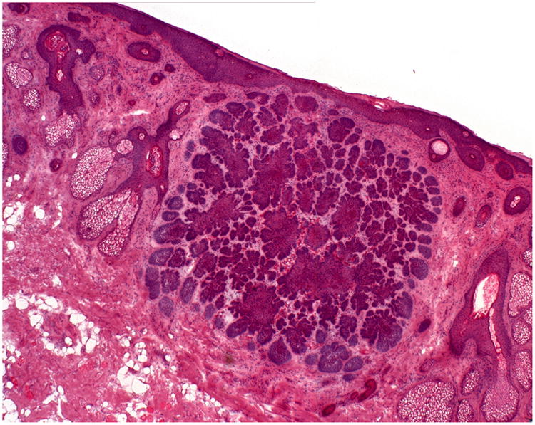
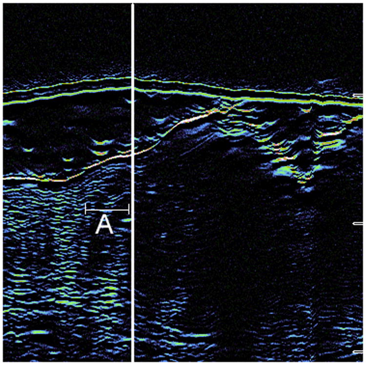
Figure 2.

A: Hematoxylin & Eosin staining from Stage I Mohs surgery slides demonstrate a large nodular tumor infiltrating the dermis. The ultrasound was able to detect this area of extension (true positive).
B: Seen here is the correlated ultrasound image from the photographed 2A case. The thick white line represents the Mohs surgeon's delineated surgical margin. The area to the left of the demarcated line (around the letter A) is the area of true positivity.

