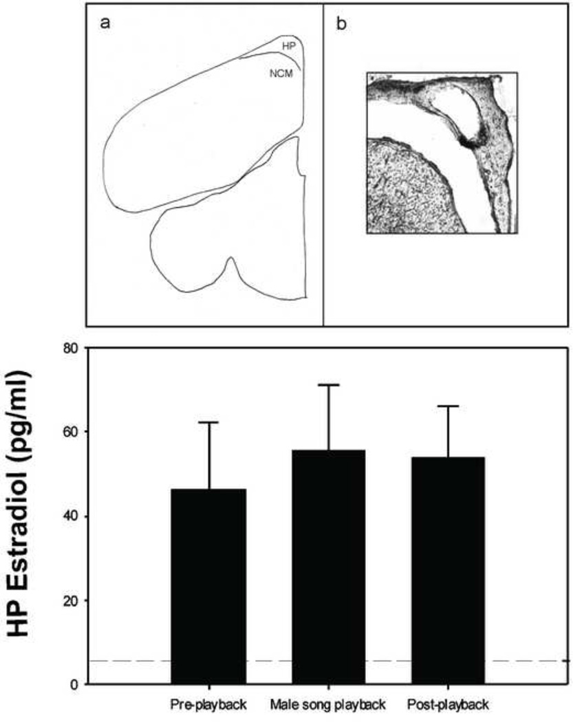
Figure 1.
Top panel: a) coronal section schematic showing location of caudal telencephalon targeted for microdialysis in the HP and NCM (figure adapted from zebra finch brain atlas; coordinates = 1.35mm rostral to the bifurcation of the midsaggital sinus (Nixdorf-Bergweiler & Bischof, 2007; Remage-Healey et al., 2008). The HP and NCM lie adjacent to one another separated by the lateral ventricle. b) Photomicrograph showing typical damage to the left HP caused by the microdialysis probe. Note the artificially enlarged distance between the HP and underlying caudal telencephalon (lateral ventricle) caused by section mounting. Bottom panel: mean HP estradiol levels (± 1 SEM) in male zebra finch HP (n= 4) for 30 minutes before, during, and after male song playback. Unlike in the adjacent NCM (Remage-Healey et al., 2008), there was no increase in HP estradiol levels in response to playback (F2,6 = 0.55; P = 0.605). Microdialysis details: during guide cannula implantation surgery, a CMA7 microdialysis probe was aimed at the following coordinates: 1.6mm rostral to the bifurcation of the midsagittal sinus and 0.5mm lateral to the midline. The guide cannula was placed on the surface of the brain and secured in place with dental cement. To begin collection, the microdialysis probe was implanted and secured with cyanoacrylate, then constantly perfused with artificial cerebral spinal fluid at a rate of 2µl/min. Individuals were housed singly in soundproof chambers with ad lib food and water throughout microdialysis sample collection. Estradiol levels were assessed in HP dialysate using the Cayman Chemical Estradiol EIA kit. For more details on microdialysis procedures, see Remage-Healey et al., 2008.

