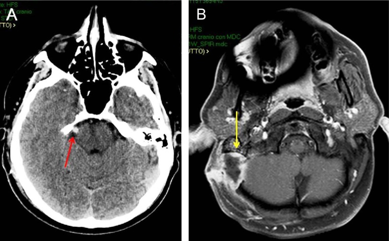
Figure 1.
Postoperative CT scan of a 40-year-old man submitted to MVD for right TN (A; red arrow). Brain axial MRI after gadolinium administration (B) 2 months after MVD, showing an abscess at the site of operation (yellow arrow).
Abbreviations: CT, computed tomography; MVD, microvascular decompression; TN, trigeminal neuralgia; MRI, magnetic resonance imaging.

