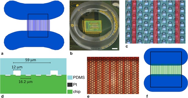
Fig 1. Geometry and layout of poly(dimethylsiloxane) axonal channel device on microelectrode array chip (a) Drawing of axonal channel device, with 12 μm channels, and neuronal culture chambers; rectangle shows dimensions of electrode array, which is 1.75 x 2.0 mm2.

(b) Photograph of packaged chip wire-bonded to a printed circuit board and with channel device on top, scale bar is 2 mm, the plastic ring is 18 mm in diameter and used to hold cell medium. (c) Zoom in on electrodes and channels: channels are lighter and highlighted in red; scale bar is 10 μm. (d) Cross section of chip, electrodes and channel device on top showing characteristic dimensions and corresponding to c. (e) Photograph of channel region of device; channels were filled with red food coloring to illustrate tightness of bonding to chip surface; scale bar is 20 μm. (f) Smaller chambers and channels used for some experiments. Channels were 2–8 μm in width; black box corresponds to array, which is 1.7 x 2.0 mm2.
