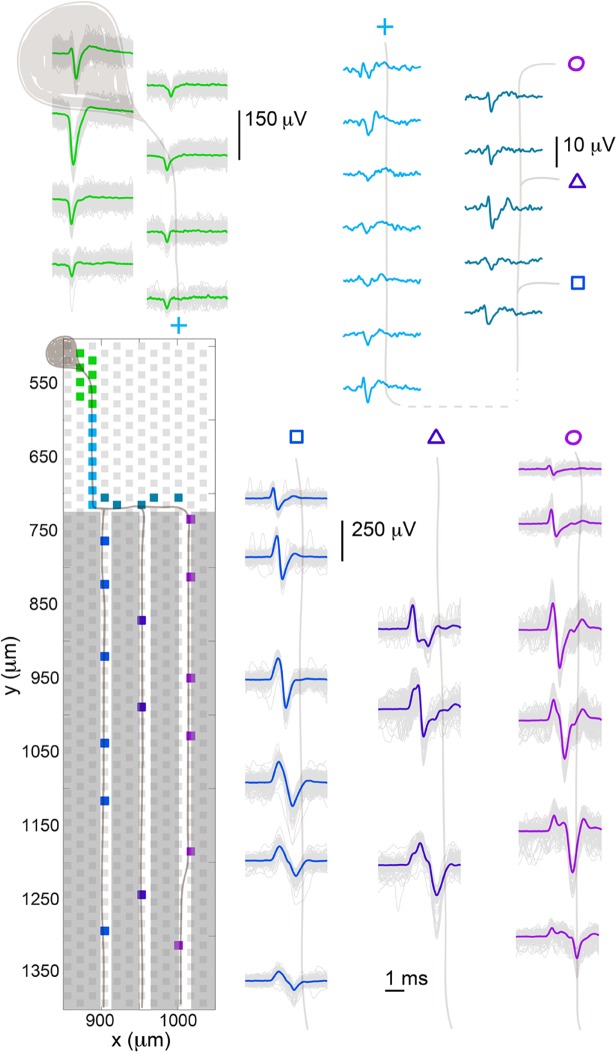
Fig 5. Spontaneous spike propagation from a soma to a branched axon.
Spike-triggered averaging of the large green somatic spike revealed a branching axon, which grew into three channels. The footprint of the soma was spread across several electrodes and showed the typical somatic shape: essentially monophasic, negative spike. The small axonal signal outside of the channels is shown in blue. Electrodes in three adjacent channels recorded spikes that were time aligned with that of the soma, and their positions and spike shapes are shown. The spike amplitudes were very different for the different cellular components (soma, axon outside channels, axon inside channels) as indicated by the scale bars. A cartoon neuron was drawn over the traces to guide the eye.

