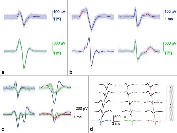
Fig 7. Spontaneous spike shapes on different electrodes in the channels during a single recording session.
Spike shapes on a given electrode were remarkably consistent and reproducible over short time periods. (a) The ‘classical’ axonal signal, which is biphasic (blue) or tri-phasic (green), starting with a positive peak, was the shape most often seen. (b) Variations on the classical shape showed either a bump before the first peak (first column) or a bump just after the spike (second column). Colored scale bars correspond to waveforms of the same color. (c) Complex multiple spike shapes in channels. A single spike shape propagated in a single direction while different shapes often propagated in opposite directions. In all cases many trials are shown and median traces are overlaid. (d) Timing of different spike shapes shown in c in the lower right plot; the positions of the electrodes are shown on the right side: the electrode of interest is light blue, and the other electrodes in the channel are outlined in black. Electrode pitch is 17 μm.

