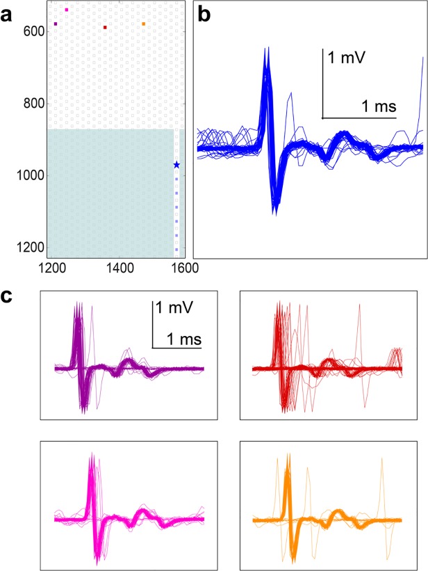
Fig 8. Complicated spike shapes originated from single cells.
Complicated spontaneous spikes could be reproduced by stimulations of a single cell at various positions. (a) Positions of electrodes that were stimulated and recorded from. The electrode of interest in the channel, nearest the entrance, is denoted with a star. (b) The spontaneous spike shape on the electrode depicted with a star in a. The large, initial, mostly biphasic spike is followed by a specific and complicated shape. (c) Stimulation at four different sites spanning 250 μm and denoted by the burgundy (left most), pink, red, and orange (right most) electrodes, all elicited the same spike at the electrode of interest in the channel. Latencies varied slightly, and jitter was introduced, but the evoked spike shape remained the same as the spontaneous spike shape.

