
Figure 2. Key Neural Networks involved in Visual and Auditory Statistical Learning.
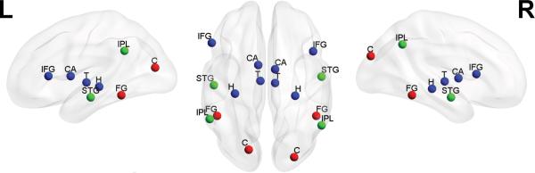
Key brain regions associated with domain-general (blue), and lower- and higher-level auditory (green) and visual (red) modality-specific processing and representation, plotted on a smoothed ICBM152 template brain. The depicted regions are not intended to constitute an exhaustive set of brain regions subserving each domain. C = Cuneus, FG = Fusiform Gyrus, STG = Superior Temporal Gyrus, IPL = Inferior Parietal Lobule, H = Hippocampus, T = Thalamus, CA = Caudate, IFG = Inferior Frontal Gyrus. Generated with the BrainNet Viewer [89].