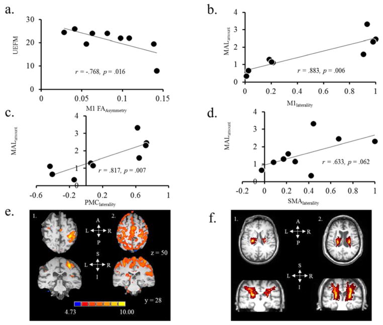
Figure 4.
Relationship between clinical assessment scores (UEFM & MAL) and neurophysiologic and neuroimaging outcomes. (a) There was a negative correlation between UEFM and M1 FAasymmetry. (b–d) there was a positive correlation between MAL and fMRI laterality index within the M1, PMC and SMA. (e) FMRI based images in two different patients. Subject (1) shows high fMRI laterality signifying good ipsilesional hemisphere dominance during contraction of the paretic hand. Subject (2) shows low fMRI laterality signifying weaker ipsilesional hemispheric dominance during contraction of the paretic hand. Their MAL scores were 2.3 and .66, respectively, signifying that patient with higher laterality showed higher MAL. (f) DTI-based images in two different patients. Subject (1) shows poor M1 FAasymmetry signifying weaker integrity of corticospinal tracts originating from the ipsilesional side. Subject (2) shows good M1 FAasymmetry signifying similar integrity of tracts in both hemispheres. Their UEFM scores were 35 and 50, respectively, signifying that patient with higher function showed favorable integrity on ipsilesional side
S = superior, I = inferior, A = anterior, P = posterior, L = left & R = right.

