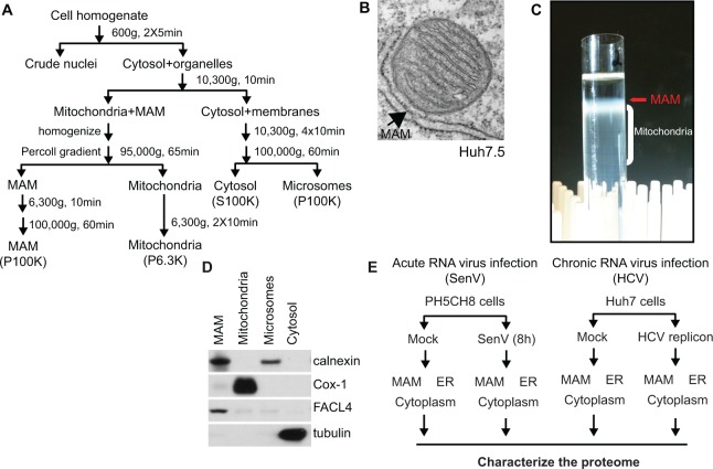
Fig 1. Experimental design for proteomic analysis of MAM fractions during RNA virus replication.
(A) Percoll gradient biochemical fractionation scheme. (B) Electron micrograph of Huh7.5 cells. MAM is indicated by the arrow pointing to the membrane wrapping around a mitochondrion. (C) Percoll gradient illustrating a representative fractionation of PH5CH8 cells at the mitochondria/MAM isolation step. MAM and mitochondria were extracted from the gradient by a needle at the location indicated by the arrow and subjected to further purification. (D) Immunoblot analysis of biochemical fractions isolated from PH5CH8 cells. Fractionation markers: calnexin, ER; Cox-1, mitochondria; FACL4, MAM; tubulin, cytosol. (E) Experimental scheme for proteomic analysis.

