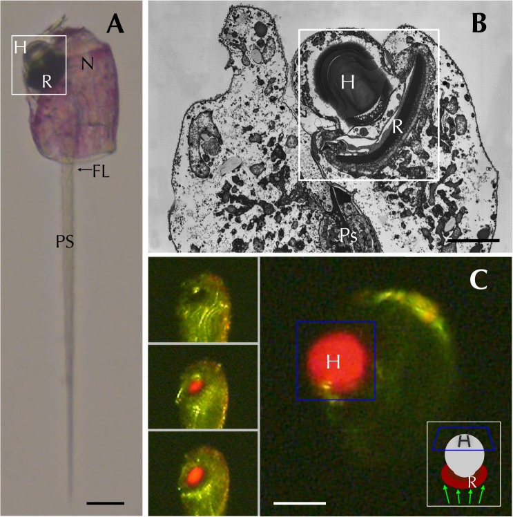
Fig 1. Erythropsidinium spp. and its subcellular structure eyespot “ocelloid”.
(A) Light micrographs (LM) of Erythropsidinium spp. H = hyalosome (crystallin body), R = retinal body, N = nucleus, FL = flagella, PS = piston. (B) Transmission electron micrographs (TEM) of ocelloid. (C) The refractile nature of the hyalosome under fluorescent microscopy. Bars: 20 μm (A, C), 10μm (B). The ocelloid is located at the left side of a cell seen in ventral view according to the orientation proposed by Kofoid and Swezy[12] (Fig. 1A). The nucleus was ellipsoidal and at the opposite side of ocelloid, in the anterior of the cell (Fig. 1B). These indices are consistent with the taxonomic criteria of the type specimen that was identified as Erythropsidinium agile. From the serial pictures of autofluorescence in the retinal body (Fig. 1C), lens-effect of the hyalosome can be observed. The front image of the retinal body is larger than side view.

