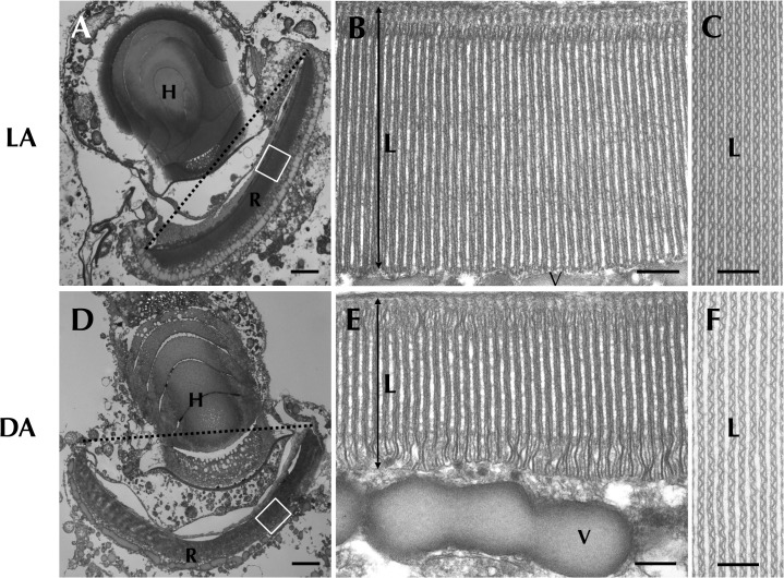
Fig 2. Retinal body of the ocelloid (R) changes its morphology in different light conditions.
Light-adapted state (LA: A, B, C) and dark-adapted state (DA: D, E, F) were observed. Enlargement of a longitudinal section of the retinal body (B, E) and cross section (C, F) are shown. L = lamellae, V = vesicular layer. Bars: 2 μm (A, D), 200 nm (B, C, E, F).

