Fig. 3.
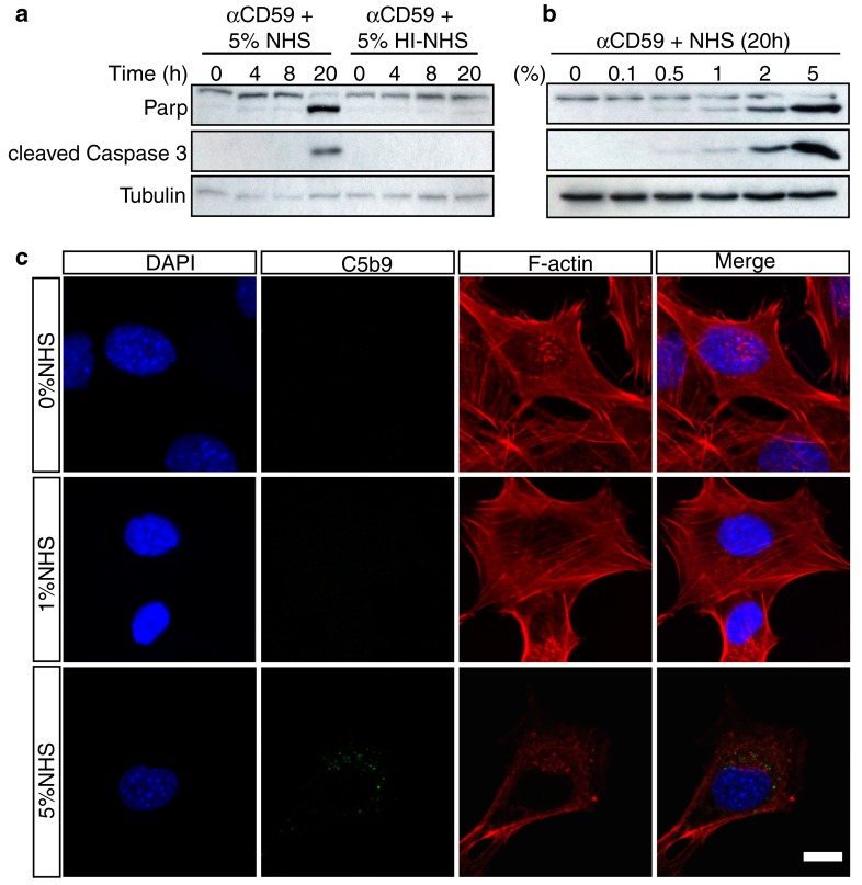
C5b-9 induces signature features of apoptosis in 661W cells in a dose- and time-dependent manner. a 661W cells were serum-starved for 24 h, then treated with anti-CD59 for 1 h followed by 5 % NHS for 1 h. Cells were lysed after 0, 4, 8 and 20 h and subjected to immunoblot analysis for PARP and cleaved caspase 3. Cells were treated with 5 % HI-NHS as a negative control and α-tubulin was included as a loading control. The blot shows that cleavage of PARP and activation of caspase-3 after 20 h occurred only in cells treated with NHS. b 661W cells were prepared as in (a), and treated with different concentrations of NHS for 20 h before immunoblotting. PARP cleavage and caspase-3 activation were faintly evident in 1 % NHS, but strikingly so in 2 and 5 % NHS. c 661W cells were prepared as in (a) with exposure to NHS for 20 h, then fixed, immunostained for C5b-9 and F-actin with nuclear counterstaining, and examined by confocal microscopy to investigate morphological changes. The images show that in 0 and 1 % NHS, 661W cells maintained a normal healthy morphology with abundant F-actin stress fibres, whereas in 5 % NHS surviving cells typically were smaller with fewer stress fibres and F-actin clumping. Scale bar 10 μm. All experiments were repeated at least three times, and representative blots/images are shown

