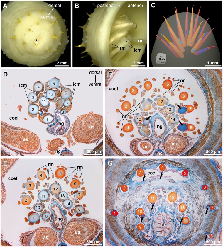
Fig 5. Echiurus echiurus, anal chaetae.
A. Extended focus view of the caudal face of the animal, B. Extended focus semi-lateral view into the dissected caudal end; Hindgut is removed, nerve cord (nc) marks ventral side. Note complex muscular system, interwoven interconnecting muscles (icm), and twisted orientation of anal chaetae. C. Reconstruction of completed (orange) and developing (bluish) anal chaetae based on a series, D-G. Azan stained histological sections from a series of transverse sections from anterior to posterior. The chaetal sacs extend deeply into the coelom (coel), the anal chaeta are numbered (outer hemi-circle 1–8, inner hemi-circle 9–14). Distances: D-E = 600 μm, E-F = 200μm, F-G = 600 μm. D. Section of the base of the anal chaetae dorsal to hindgut. Note connection of chaetal sacs by interconnecting muscles (icm). E. Section of the chaetae at the level of the origin of the radial muscles (rm). Anal chaetae are grouped in two hemi-circles that are next to each other and partly surround the hindgut (hg). F. Section close to the posterior end of the trunk body cavity (coel). Hemi-circles of anal chaetae are clearly separated and surround three quarters of the hindgut. Note developing chaeta (arrow) in the sacs of chaetae 7, 8, and 10. G. Section of the anal chaeta through the body wall muscles. Hemi-circles of anal chaetae are widely separated. Note tips of developing chaetae (arrow) in sacs of chaetae 3, 4, 7, 8, 10, and 11. a anus, as anal sac.

