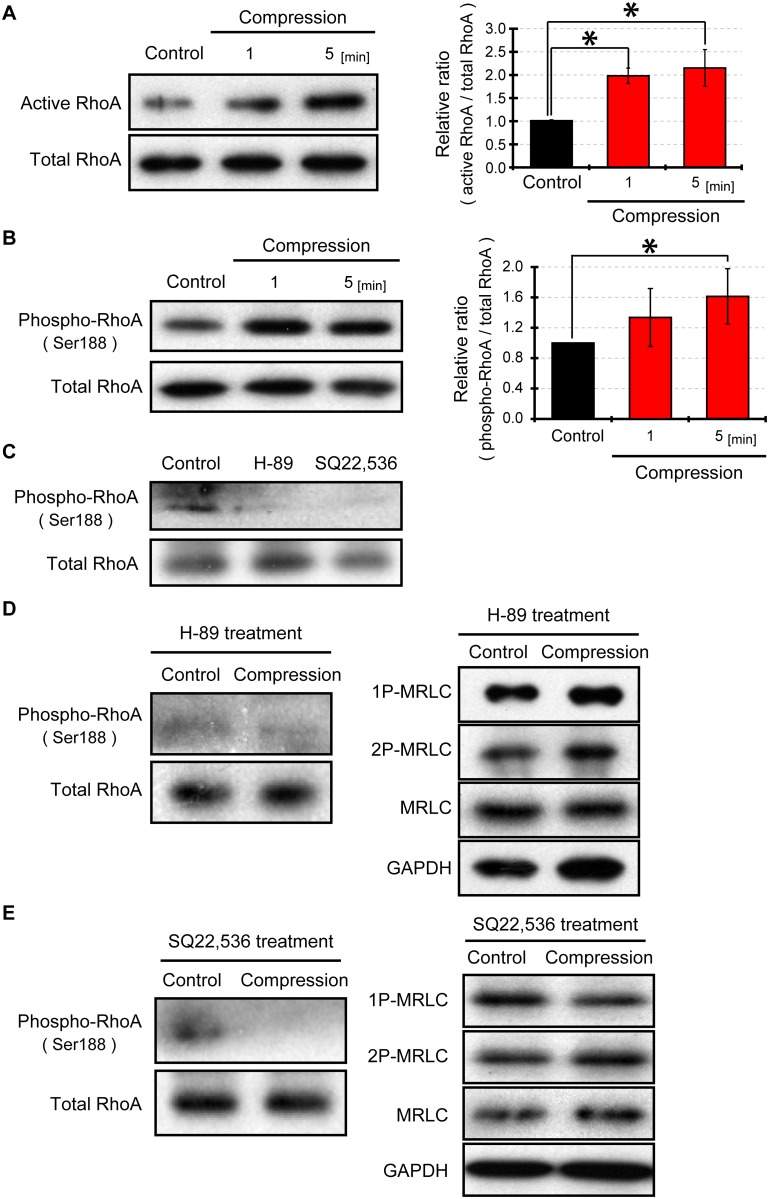
Fig 5. Compression induced RhoA activation and phosphorylation.
(A) Detection of active RhoA under compressive loading. Active RhoA (RhoA-GTP) was precipitated with Rhotekin-RBD beads. Total RhoA was used as a loading control. (B) Western blotting for phosphorylated RhoA after compressing cells. RhoA (Ser188) phosphorylation was detected by western blotting, and total RhoA was used as a loading control. (C) Effects of protein kinase A (PKA) inhibitor H-89 or adenylyl cyclase inhibitor SQ22,536 on phosphorylated RhoA (Ser188). Cells on a plastic dish were treated with 30 μM H-89 or 100 μM SQ22,536 for 90 minutes. (D) Influence of PKA inhibition on compression-induced RhoA phoshorylation and MRLC dephosphorylation. Cells were compressed under the presence of 30 μM PKA inhibitor H-89. (E) Effect of inhibition of adenylyl cyclase on compression-stimulated RhoA phoshorylation and MRLC dephosphorylation. Cells were treated with 100 μM adenylyl cyclase inhibitor (SQ22,536) for 90 minutes before cell compression. The levels of active RhoA, phospho-RhoA, total RhoA, 1P-MRLC, 2P-MRLC, MRLC and GAPDH were detected by western blotting and quantified with Image J software. Quantification of western blots represents an average of 3 independent experiments, mean ± SEM. *, p < 0.05.

