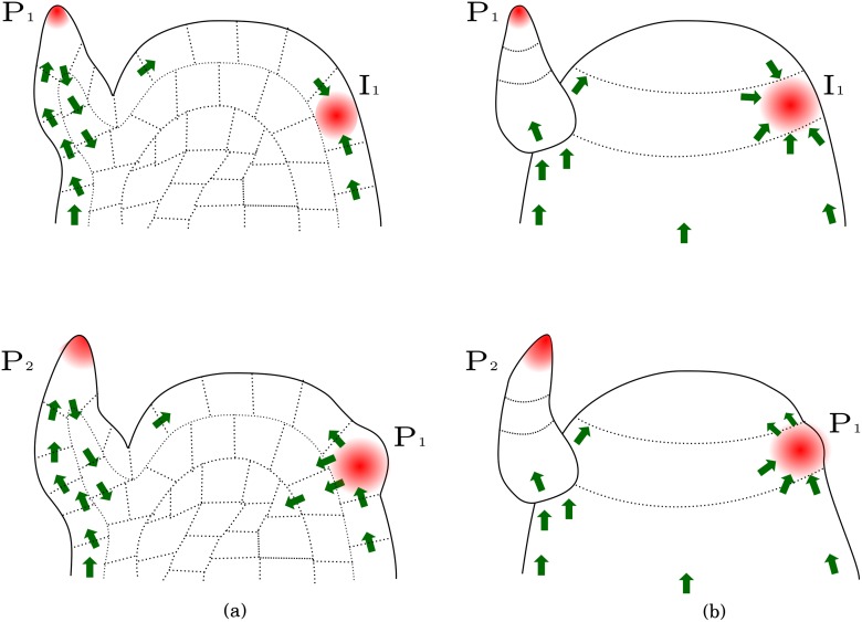
Fig 1. Schematic representation of a shoot apical meristem showing auxin canalization.
(a) Transversal section. The upper panel shows PIN polarization (green arrows) soon after incipient primordium formation (I 1), and the lower panel shows a later stage. (b) External view of the same meristem and developmental stages. PIN proteins are initially polarized towards auxin maxima close to primordia (upper panel). Notice that PIN polarization is later reversed in adaxial cells (lower panel).

