Figure 3.
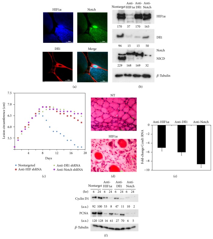
HIF-Dll-Notch axis in pcRNA-induced myogenesis. (a) A triple-stained section of injured quadriceps muscle treated with pcRNA1–3 combination for 4.5 h, showing HIF1α (blue) concentrated in the nucleus of an activated satellite cell, colocalizing with Notch (green) and Dll1 (red) attached to the plasma membrane as well as the plasma lemma of the underlying myofiber. Scale bar, 10 μm. (b) Effect of RNA interference on protein expression in injured muscle infected for 2 d with lentivirus expressing shRNA targeted against the indicated mRNAs and then administered pcRNA1–3 for 6 or 24 h, before western analysis of indicated proteins. (c) Effect of knockdown of the indicated proteins on wound resolution. (d) Hematoxylin-eosin stain of lentivirus-infected injured muscle expressing nontargeted (NT, upper) or HIF1α-targeted shRNA (lower), treated with pcRNA for 2 weeks. Scale bar, 100 μm. (e) Fold-change in cyclin D1 (Ccnd1) mRNA level, relative to nontargeted control, in injured muscle knocked down for the indicated protein and treated with pcRNA for 6 h, determined by Q-PCR. (f) Western blots of nontargeted or targeted muscle treated with pcRNA1–3 for 6 or 24 h, probed for Ccnd1, PCNA, or β-tubulin.

