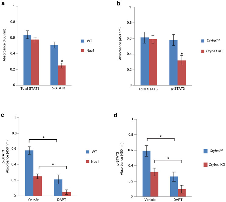
Figure 2. p-STAT-3 is impaired in astrocytes lacking βA3/A1-crystallin.
(a). Analysis of WT and Nuc1 astrocyte lysates with total STAT3 and p-STAT3 antibodies using sandwich ELISA. No difference in the level of total STAT3 was observed between WT and Nuc1 astrocytes, whereas, the level of p-STAT3 was significantly reduced (~50%) in Nuc1 astrocytes compared to WT astrocytes. (b). Cryba1 KD mouse astrocytes also showed significant reduction in the level of p-STAT3 compared to the Cryba1fl/fl controls. Levels of total STAT-3 were unchanged. (c). Both WT (~57%) and Nuc1 (~75%) rat astrocytes treated with DAPT showed significant reduction in p-STAT3 compared to vehicle-treated cells. (d). Likewise, Cryba1fl/fl and Cryba1 KD astrocytes showed ~ 50% and ~62% reduction in p-STAT3 compared to respective vehicle-treated cells. Error bars indicate s.d.; *P<0.05.

