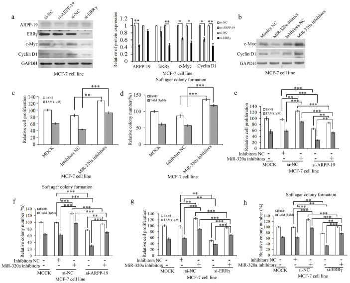
Figure 2. Knockdown of miR-320a reduced sensitivity of tamoxifen in MCF-7 cells.
(a). Western blot analysis of ARPP-19, ERRγ, c-Myc, and Cyclin D1 expression levels in MCF-7 cells transfected with si-ARPP-19 and si-ERRγ and the corresponding densitometric analysis. (b). Western blot analysis of c-Myc and Cyclin D1 expression levels in MCF-7 cells transfected with miR-320a mimics/inhibitors. c and d. Cell viability (c) and soft agar colony formation (d) analysis of MCF-7 cells transfected with miR-320a inhibitors. e and f. Cell viability (e) and soft agar colony formation (f) analysis of MCF-7 cells transfected with si-ARPP-19 and miR-320a inhibitors and co-transfected with si-ARPP-19 and miR-320a inhibitors on exposure to tamoxifen or not. f and g. Cell viability (f) and soft agar colony formation (g) analysis of MCF-7 cells transfected with si-ERRγ and miR-320a inhibitors and co-transfected with si-ERRγ and miR-320a inhibitors on exposure to tamoxifen or not. Results are presented as an average of at least three replicates. *P < 0.05, **P < 0.01, ***P < 0.001.

