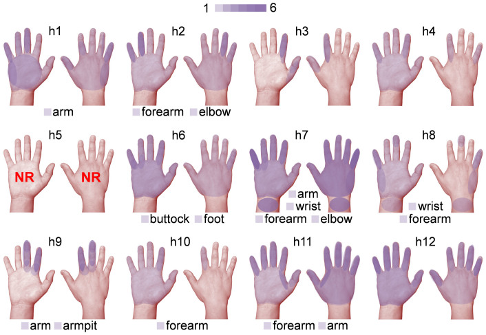
Figure 2. Illustration depicting the locations of tactile sensations experienced by the subjects under FUS stimulation.
The regions of sensations felt from the left and right hands, including the wrist, as represented by the semi-transparent purple layers, were merged onto the palmar (left) and dorsal (right) view of the right hand (‘h1' through ‘h12'). The number of occurrences for a set of distinctive locations of sensation are represented by a color scale (1–6). The locations of other reported sensations (i.e. arm, forearm, armpit, elbow, wrist, buttock, and foot) were labeled at the bottom of each panel. Subject ‘h5' did not report any sensations (noted as ‘NR').

