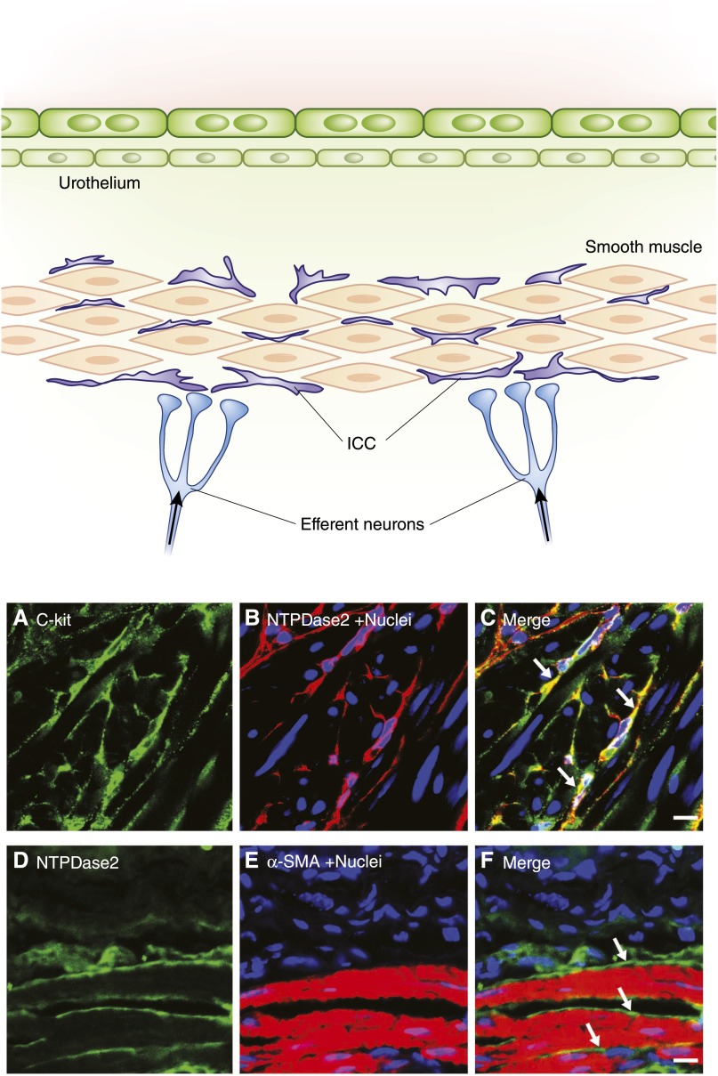
Figure 9.
Morphology and location of BICCs. The top panel provides a schematic of the bladder wall showing the location of ICCs adjacent to smooth muscle bundles and parasympathetic neurons. These stellate-shaped cells are also seen in the deep lamina propria. The lower panels show immunofluorescence staining of BICCs in 5-µm cryosections of BSM from mouse. (A) c-kit Staining. (B) Ectonucleotidase2 (NTPDase2) staining. (C) Merge showing colocalization (in yellow) of the two BICC markers (arrows). (D–F) images showing close apposition of BICC (arrows) and BSM cells as indicated by NTPDase2 and α-smooth muscle actin. α-SMA, α-smooth muscle actin; BICC, bladder interstitial cell of Cajal; BSM, bladder smooth muscle; ICC, interstitial cell of Cajal. Bar, 10 µm.

