Figure 1.

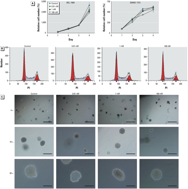
Long-term exposure to B[a]P showed no detectable effects on HCC cell growth. (A) BEL-7404 (left) and SMMC-7721 (right) cells were treated with 0.1% DMSO or different concentrations of B[a]P for 1 month, and cell growth was evaluated by the CCK-8 assay; values are mean ± SD (n = 6/group). (B) BEL-7404 cells were treated with 0.1% DMSO or B[a]P for 1 month and harvested for cell cycle distribution analysis by flow cytometry. Red areas represent G1/S and G2/M phases; arrowheads indicate the peaks of G1/S and G2/M phases. (C) Soft agar assay of BEL-7404 cells exposed to 0.1% DMSO or different concentrations of B[a]P for 1 month. Bars = 1 mm for 4× magnification (top), 400 μm for 10× magnification (center), and 200 μm for 20× magnification (bottom).
