
Figure 3.
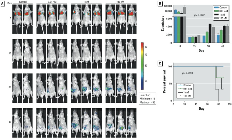
B[a]P-exposed HCC cells metastasized more extensively in nude mice than did control cells. (A) Monitoring of metastasis of bioluminescent SMMC-7721 cells exposed to B[a]P; images were obtained at 30 min (0 days), 15 days, 30 days, and 40 days after injection with cells. (B) Quantification of photon counts for each group of mice over time; values are mean ± SD (n = 2–3/group; p = 0.0032 by two-way ANOVA). (C) Kaplan-Meier overall survival curves for mice injected with HCC cells exposed to different doses of B[a]P; B[a]P treatment reduced the overall survival rate in a concentration-dependent manner (p = 0.0159, log-rank test for trend).