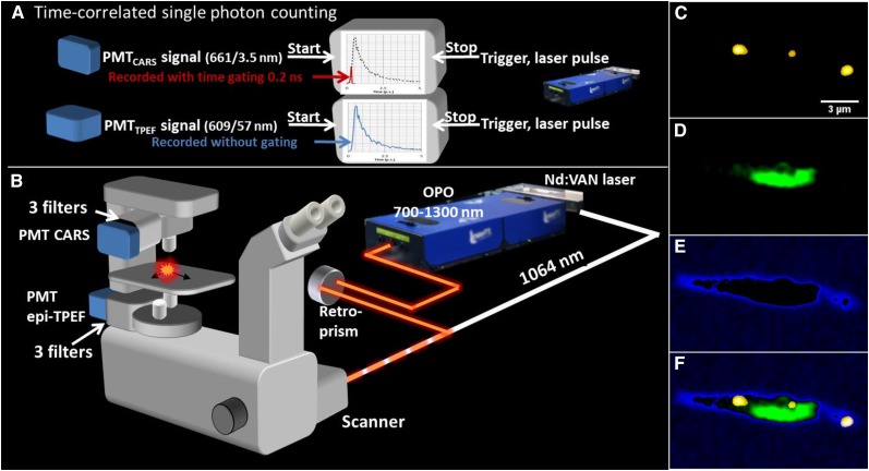
Figure 1.
A, Schematic illustration of the time-gated CARS microscope allowing efficient separation of TPE fluorescence from chloroplasts in algae/plant cells using time-correlated single-photon counting. A neodymium vanadate pump laser generates the Stokes beam (1,064 nm) and seeds the optical parametric oscillator (OPO). The Stokes and pump/probe (typically at 816.9 nm) pulse trains are overlapped in time and space and coupled into the inverted microscope through a mirror scanner. Signals are recorded by single-photon counting photomultiplier tubes (PMTs) after spectral filtering (CARS in transmission mode, TPE fluorescence in epi mode) and processed by time-correlated single-photon counting. The instantly emitted CARS photons (spectrally filtered at 665/7.5 nm) are recorded during a gating time of 200 ps (red), whereas the full time decay is recorded for the TPE fluorescence (filtered at 684/24 nm; blue). The decay curve is symbolic. C to F, Spectrally filtered and gated images of algae grown for 159 h under Ctrl conditions. Shown are a CARS microscopy image (C-H vibration of 2,845 cm−1) of intracellular lipid droplets (yellow; C), TPE fluorescence of chloroplast (green; D), thresholded low-intensity CARS signal from the cell soma illustrating the outline of the cell (blue; E), and the overlay (F) of images C to F illustrating the location of the lipid droplets throughout the cell soma and in relation to the chloroplast.

