
Figure 2.
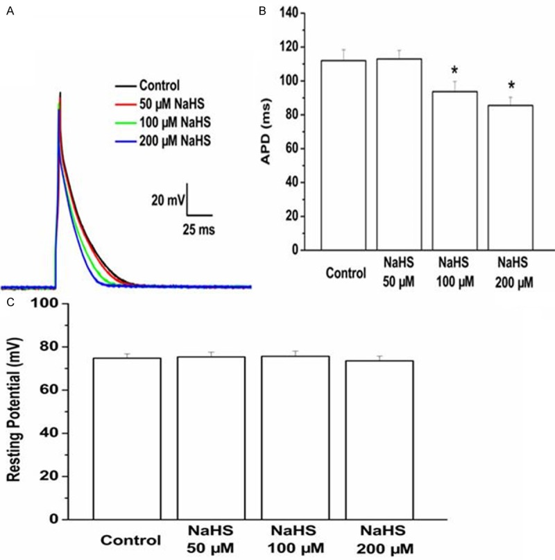
Effect of exogenous NaHS on the action potential duration and resting potential cardiomyocytes from myocardium around ischemic area were isolated and treated with NaHS. The NaHS levels were 50, 100, 200 μM. The action potential duration (A, B) and resting potential (C) were tested by patch-clamp amplifier. Data are the mean ± SD of six cells. *P < 0.05 vs control group.