
Figure 2.
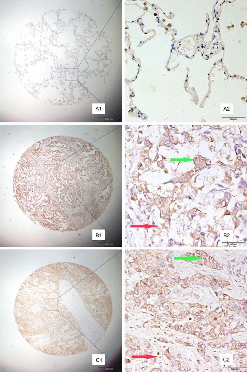
Representative patterns of MAGE-A9 protein expression showed in non-small cell lung cancer (NSCLC) and adjacent noncancerous tissues. Case A, the adjacent tissue, shows negative staining. Case B, the lung adenocarcinoma tissue, presents high expression level of MAGE-A9 (green arrow). The stromal tissue was also detected MAGE-A9expression (red arrow). Case C, lung squamous cell carcinoma tissue, shows strong MAGE-A9 protein expression in both cancer cells (green arrow) and stomal cells (red arrow). Original magnification: (A1-C1) × 40, (A2-C2) × 400.